
PROCTOLOGY
Pilonidal Sinus
A common disease of the natal cleft that can now be treated in different ways
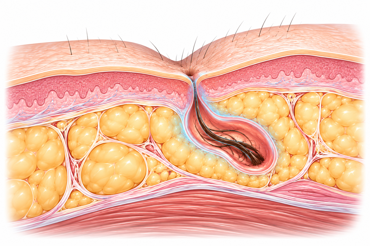
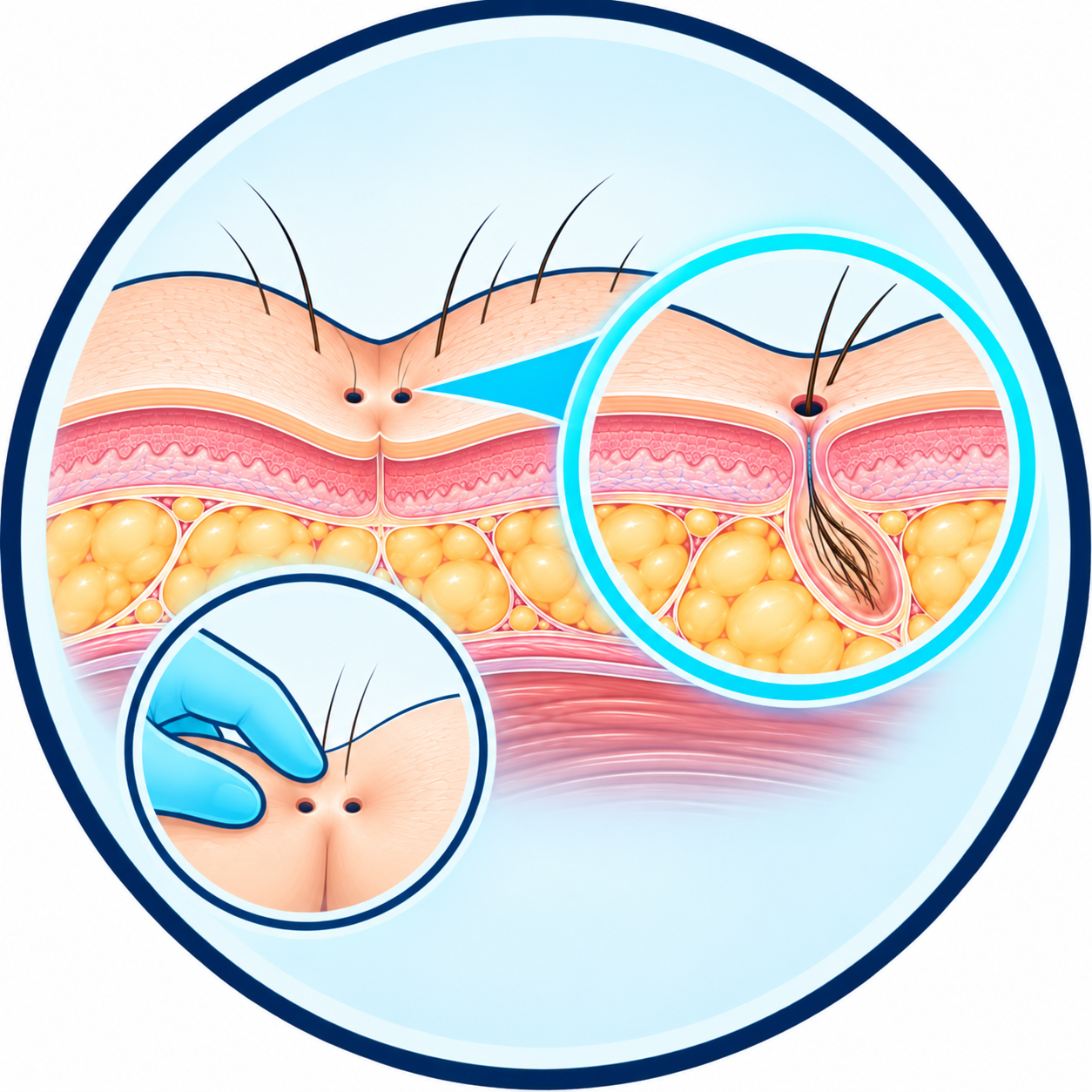
Pilonidal sinus is an inflammatory condition most often located in the natal cleft, usually a few centimetres above the anus.
Small skin openings can communicate with a cavity or tracts beneath the skin into which hairs and debris can penetrate.
The disease can remain discreet, cause repeated discharge, or present suddenly as a painful abscess.
Treatment depends mainly on its presentation and extent. Limited disease does not necessarily require the same intervention as a complex or recurrent sinus.
What is a pilonidal sinus?
An acquired condition of the skin and subcutaneous tissue
Pilonidal sinus generally develops in the upper part of the natal cleft. It can involve:
- one or more small skin openings
- a cavity located beneath the skin
- one or more secondary tracts
- trapped hairs
- a local inflammatory reaction
The term “pilonidal cyst” is frequently used, but it is not always a true closed cyst. The term pilonidal sinus or pilonidal disease often describes the condition more accurately.
How does a pilonidal sinus form?
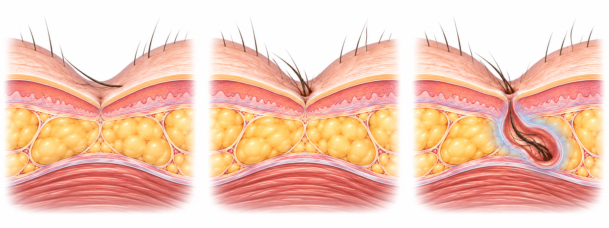
Hair, friction and skin penetration
Pilonidal disease is now considered mainly to be an acquired condition.
The depth of the natal cleft, local movement, friction and detached hairs favour their accumulation and subsequent penetration through small skin openings.
The body reacts to these hairs as foreign bodies, and inflammation can progressively form a small cavity or subcutaneous tracts.
It is therefore not simply a matter of “removing a cyst”: one must understand where the openings are and how far the disease really extends.
What are the symptoms?
The presentation can vary widely
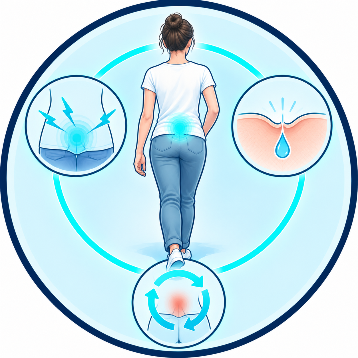
Some people simply discover one or more small openings in the natal cleft. When the disease becomes symptomatic, it can cause:
- local pain or tenderness
- a small swelling
- intermittent discharge
- repeated inflammatory episodes
- or a very painful acute abscess
Asymptomatic pilonidal disease does not automatically require preventive intervention: 2024 European guidelines advise against prophylactic surgery in asymptomatic patients.
When an abscess forms
An acute complication that usually needs to be drained
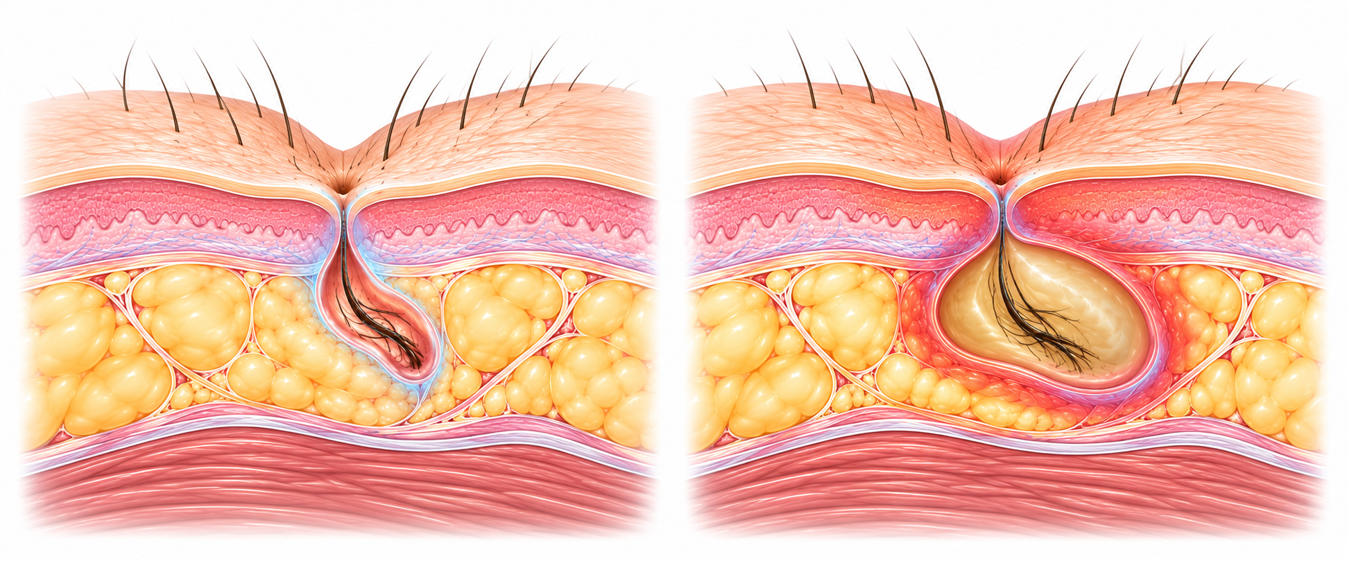
A pilonidal sinus can become infected and form an abscess. The area then rapidly becomes painful, tense, swollen and inflamed.
In this situation, the immediate goal is usually not to perform major definitive surgery: the priority is to properly drain the abscess to evacuate the collection and relieve the inflammation.
ESCP guidelines recommend incision and drainage of the acute abscess, ideally through a lateral incision. Definitive treatment of the residual disease can then be discussed.
How is the diagnosis made?
Clinical examination is usually sufficient
Diagnosis is mainly clinical. Examination looks for:
- the openings located in the cleft
- a possible secondary opening
- a swelling
- discharge
- an abscess
- scars from previous interventions
Above all, it allows the extent of the disease to be estimated. Ultrasound, MRI or other tests are usually not necessary in a typical presentation, but may be useful in certain atypical situations. An opening unusually close to the anus should in particular raise the differential diagnosis of an anal fistula.
Not all pilonidal disease looks the same
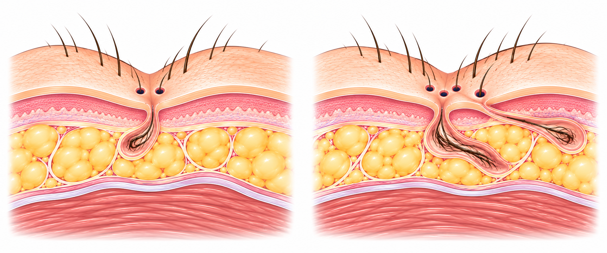
Limited disease is not the same as complex disease
This is probably the most important message on this page.
In some patients, the disease is limited to a few small midline openings and a short tract. In others, there is:
- several tracts
- lateral extensions
- repeated abscesses
- scars from earlier interventions
- or more extensive recurrent disease
This anatomical difference directly influences the choice of treatment. European guidelines allow minimal interventions to be considered for certain simple, limited forms of disease, while reserving more extensive interventions for the forms that require them.
Is surgery always necessary?
No
The incidental discovery of a few pilonidal openings without pain, inflammation or discharge does not necessarily mean that surgery is required.
Symptomatic disease, however, deserves an evaluation to determine the appropriate strategy.
Treatment can then range from a very limited procedure to reconstructive surgery, depending on the anatomy.
The right treatment is not necessarily the one that removes the most tissue. It is the one that correctly addresses the real extent of the disease.
Minimally invasive techniques
Treating limited disease while preserving as much tissue as possible
Some simple forms can be treated with limited procedures.
The principle is to treat the responsible openings and pathological tracts without performing wide excision of the whole region.
These approaches include in particular:
- pit-picking
- certain endoscopic procedures
- other selected minimally invasive techniques
These methods have the potential benefit of reducing the size of the wound and allowing a faster recovery.
They are not, however, suitable for every anatomy.
The level of evidence for some of these techniques remains limited, and their recurrence risk must be part of the discussion.
Pit-picking
Treating the small responsible openings
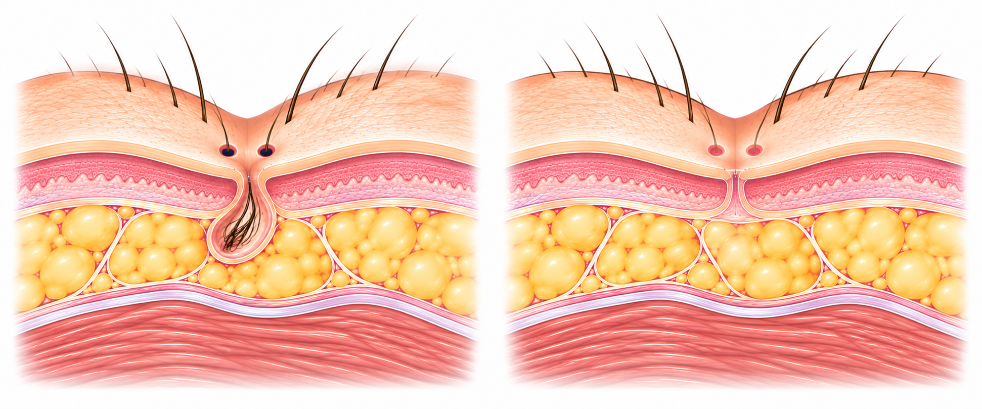
Pit-picking is a minimally invasive approach intended mainly for certain limited, non-complex forms of disease.
The principle is to remove or open, very locally, the small midline openings that sustain the disease, and to treat the associated tract or cavity with reduced tissue damage.
The goal is to avoid a large wound when the anatomical extent does not justify one.
ESCP guidelines consider that this technique can be considered when the disease is confined to the pits, possibly with a simple lateral cavity.
This does not mean it is the best choice for every case of pilonidal disease.
Endoscopic treatment — EPSiT
Seeing and treating the tract from within
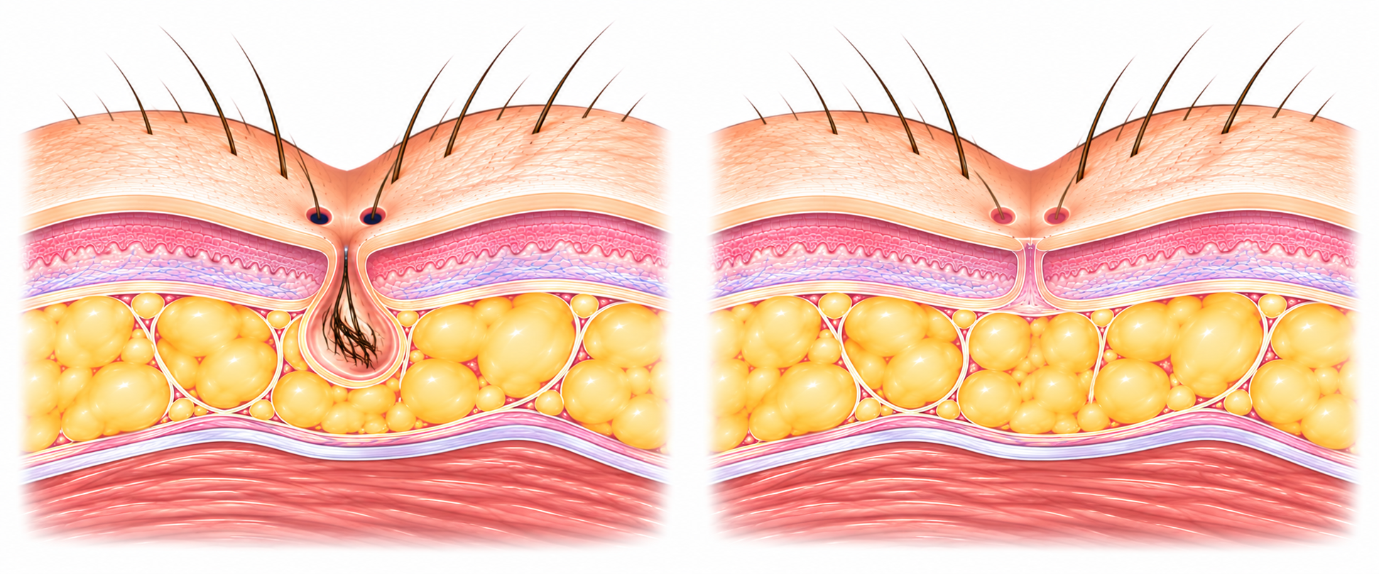
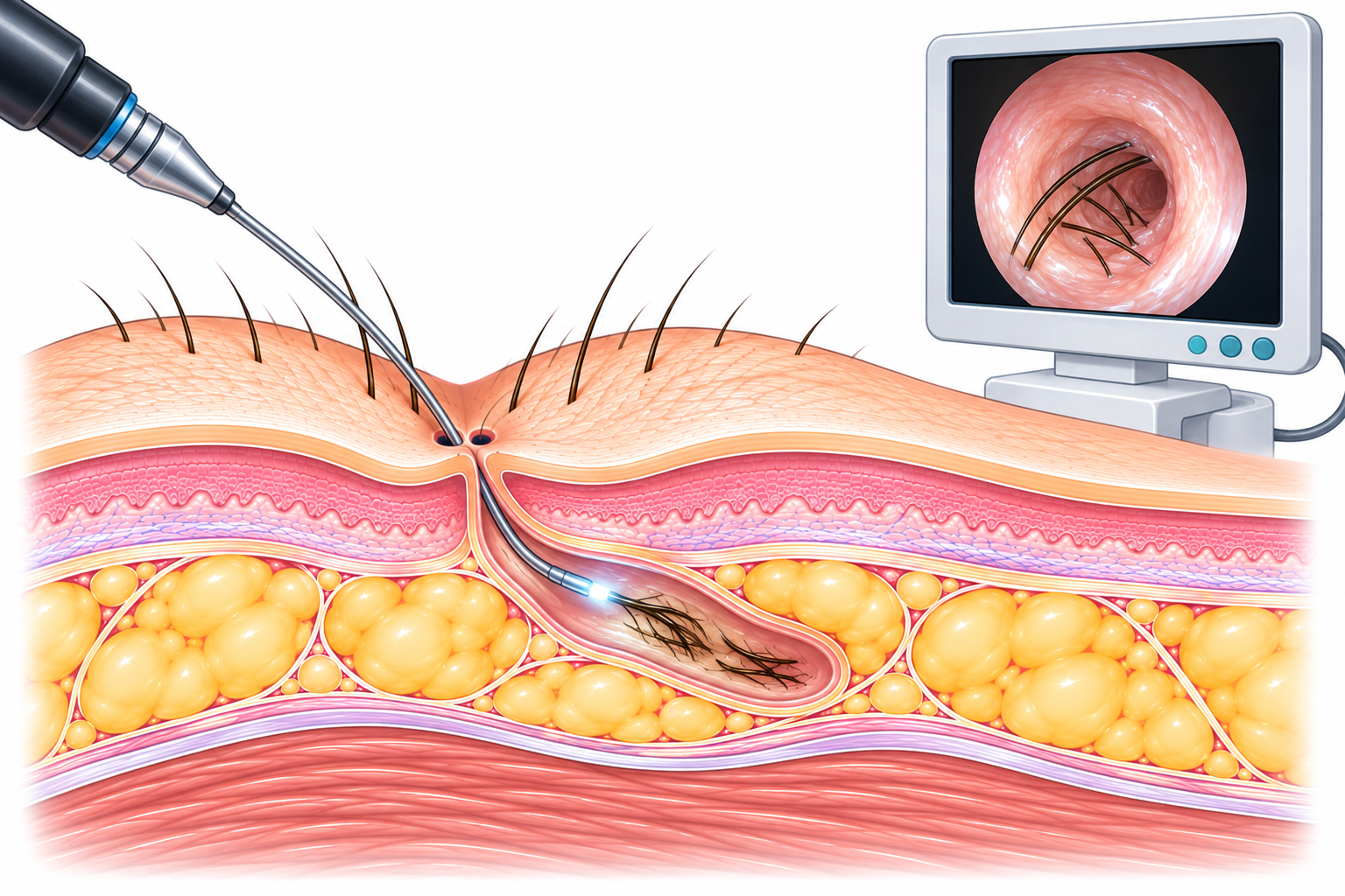
Another approach involves introducing a small endoscope into the pilonidal tract.
This technique is often referred to by the acronym EPSiT — Endoscopic Pilonidal Sinus Treatment.
It allows direct visualization of the inside of the cavity and tracts.
Hair and debris can be removed, and the pathological tissue of the tract is then treated under visual control.
The conceptual benefit is clear: treating the sinus from within while limiting the skin wound.
European guidelines consider endoscopic treatment a possible option. The level of evidence, however, is less robust than for some recommendations concerning excisional surgery.
When the disease is more extensive
The same logic, adapted to more complex anatomy
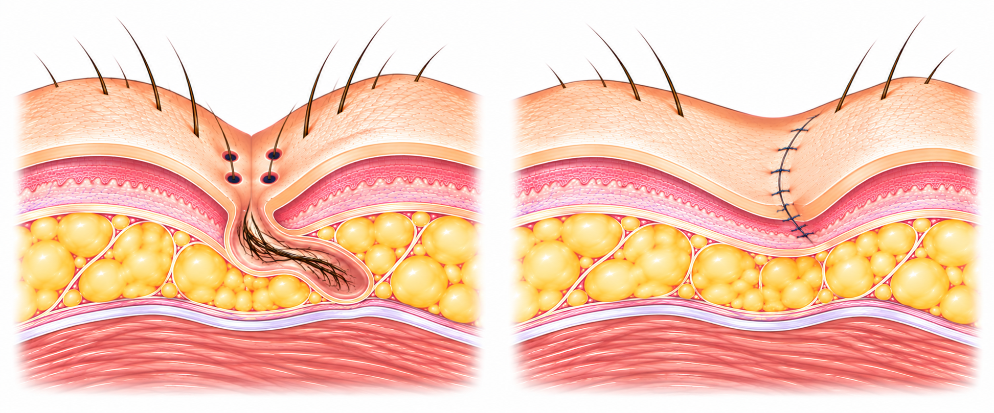
When the disease is more extensive — longer tracts, lateral extension, recurrence after a first treatment — surgery sometimes needs to remove a greater amount of pathological tissue.
In these situations, how the wound is closed becomes an important question.
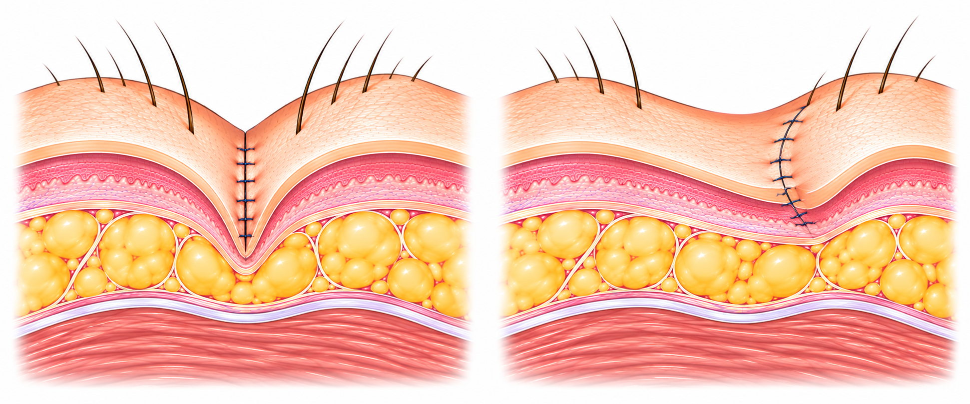
A closure made strictly along the deep midline can, in some patients, favour more difficult healing and a higher risk of recurrence, due to the tension and moisture inherent to this anatomical groove.
This is why many teams propose, for extensive or recurrent forms, closure techniques that move the scar away from the deep midline.
This is not a question of a good technique versus a bad one. It is about adapting the reconstruction to the real extent of the disease.
Off-midline reconstructions
Moving the scar to improve healing
Several techniques allow the wound to be closed away from the deep midline, notably Karydakis-type flaps or the Bascom cleft-lift, and other skin flap variants.
The common principle is to reconstruct the region so as to progressively flatten the natal cleft and position the scar away from the depth of this groove.
In the literature, these techniques are associated with lower recurrence rates than midline closure for extensive or complex disease.
They do, however, require specific surgical expertise and a recovery period that must be tailored individually.
An individualized strategy
Choosing the right option for the right anatomy
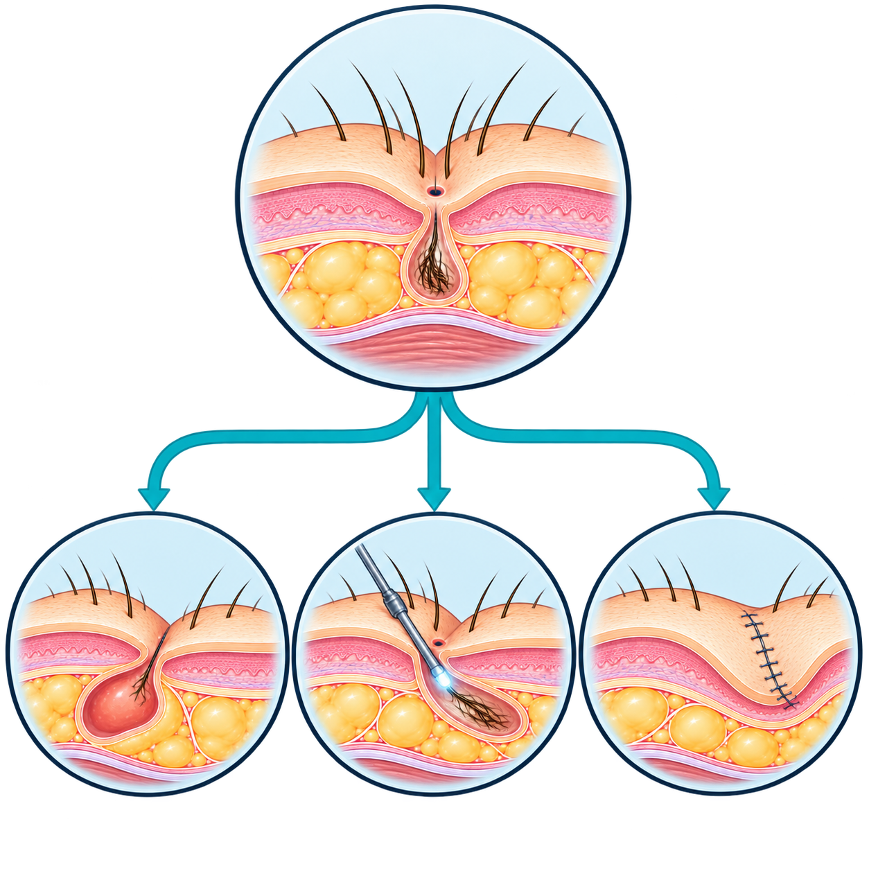
There is no single treatment for pilonidal sinus, but rather a range of techniques, each suited to a particular presentation of the disease.
The choice depends on several factors:
- the anatomical extent of the disease (number of openings, length of tracts, lateral extension)
- whether the condition is simple, complex or recurrent
- the history of treatments already performed
- the patient’s preferences and lifestyle
A precise clinical evaluation — supplemented if necessary by imaging — allows the most appropriate strategy to be proposed, from the most limited procedure to the most complete reconstruction.
After the procedure
Recovery that varies with the technique
Recovery depends directly on the procedure performed.
After a minimally invasive technique (pit-picking, endoscopy), discomfort is usually moderate and the return to activities faster.
After wider excisional surgery or reconstruction, wound care is more significant and recovery longer, with precise instructions regarding local hygiene, hair removal in the area, and the gradual resumption of activities.
In all cases, personalized postoperative instructions are given to promote optimal healing.

Can the risk of recurrence be reduced?
Simple measures that are useful alongside treatment
Certain hygiene and lifestyle measures can help reduce the risk of recurrence, without eliminating it completely:
- regular hair removal in the natal cleft area
- careful local hygiene
- maintaining an appropriate weight
- avoiding prolonged sitting when possible during the healing period
These measures are in addition to surgical treatment: they do not replace it.
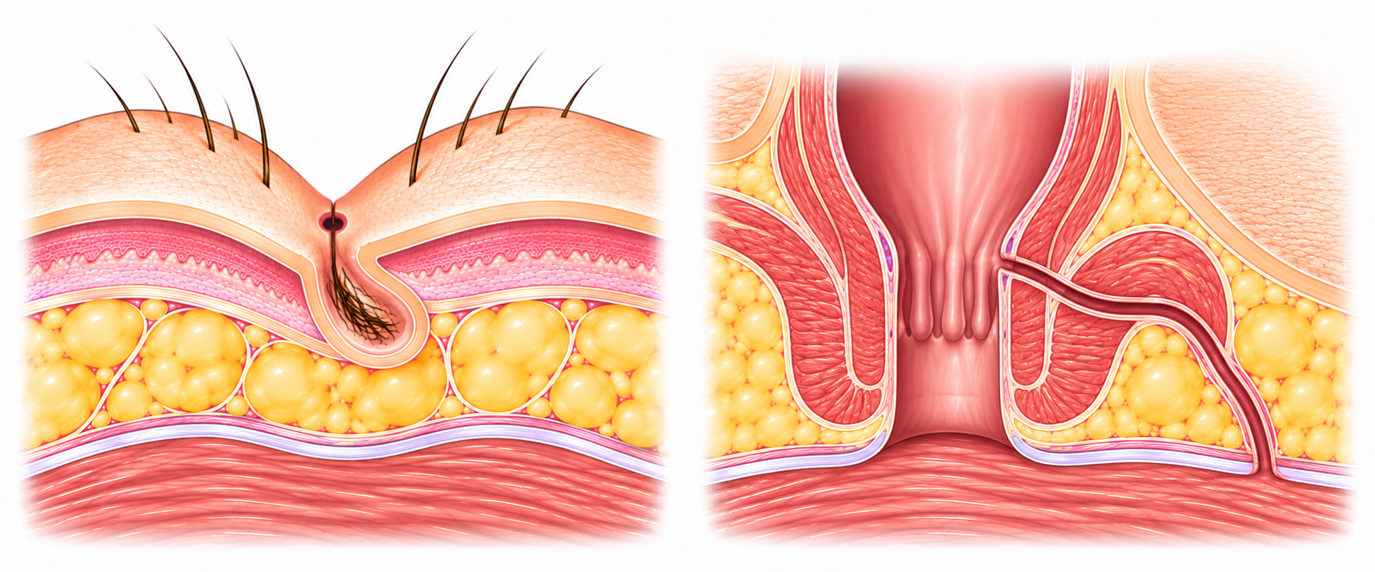
Pilonidal sinus or anal fistula?
Two different conditions, sometimes confused
Pilonidal sinus and anal fistula are two distinct conditions, although both can present with discharge or an abscess in the perineal region.
Pilonidal sinus develops in the subcutaneous tissue of the natal cleft, with no communication with the anal canal or rectum.
An anal fistula, by contrast, originates from an anal gland and communicates with the anal canal.
This anatomical distinction is essential, as it determines different diagnostic and therapeutic approaches.
In case of doubt, careful clinical examination usually allows the two situations to be distinguished.
When to seek consultation?
A consultation is recommended in the presence of:
- persistent pain or discomfort in the natal cleft
- discharge, oozing or bleeding in the cleft
- swelling, redness or local warmth suggesting an abscess
- fever associated with pain in the sacrococcygeal region
- visible opening(s) or small hole(s) in the natal cleft
- recurrence of symptoms after a previous treatment
- discomfort with prolonged sitting
- concern or diagnostic doubt, particularly regarding an anal fistula
Surgery tailored to the real extent of the disease
Pilonidal sinus covers highly variable presentations, from a simple asymptomatic opening to complex, recurrent disease.
The right treatment is not the one that systematically removes the most tissue: it is the one that precisely matches the real extent of the disease.
This individualized approach, from minimally invasive procedures to off-midline reconstruction, allows each patient to be offered the strategy best suited to their anatomy and lifestyle.

Dr Pierre Fournier
Visceral Surgeon — Proctology
Frequently asked questions
No. Pilonidal sinus results from a mechanical process involving hair penetrating the skin of the natal cleft, favoured by friction and the anatomical shape of the cleft. It is not a consequence of poor hygiene.
Yes, it is a relatively common condition, affecting mainly young men, often with dense body hair, between puberty and their thirties.
No. Disease discovered incidentally, without symptoms, does not systematically require an intervention. Symptomatic disease, however, deserves an evaluation to determine the appropriate strategy.
A simple sinus has few openings and a limited tract. A complex sinus has several openings, longer tracts, lateral extension, or a history of recurrence, which influences the choice of treatment.
Pit-picking is a minimally invasive technique that involves removing or opening, on a very local basis, the small midline openings responsible for the disease, with reduced tissue damage. It is aimed mainly at limited, non-complex forms.
EPSiT (Endoscopic Pilonidal Sinus Treatment) is an endoscopic technique that allows the pilonidal tract to be visualized and treated from within, through a small access point, while limiting the skin wound.
A strictly midline closure can, in some patients, favour more difficult healing and a higher risk of recurrence. Moving the scar away from the deep midline improves healing for extensive or recurrent disease.
These are reconstructive techniques that move the scar away from the deep midline and progressively flatten the natal cleft. They are associated with lower recurrence rates for extensive or complex disease.
It depends on the procedure performed: a few days to a few weeks after a minimally invasive technique, longer after wide excisional surgery or reconstruction, with wound care to be continued according to the instructions given.
Yes, a risk of recurrence exists, which varies depending on the technique used and the initial extent of the disease. Certain hygiene and lifestyle measures can help reduce it.
Regular hair removal in the natal cleft area, careful local hygiene, maintaining an appropriate weight, and avoiding prolonged sitting during the healing period can all help.
Yes. A pilonidal abscess presents as painful swelling, redness and sometimes fever. It often requires urgent drainage before a definitive treatment can be considered at a later stage.
No. Pilonidal sinus develops in the subcutaneous tissue of the natal cleft with no communication with the anal canal, whereas an anal fistula originates from an anal gland and communicates with the anal canal.
Yes, the European Society of Coloproctology (ESCP) has published guidelines describing the different treatment options according to the extent and complexity of the disease.
Consultation is recommended in cases of pain, discharge, swelling suggesting an abscess, associated fever, visible openings in the natal cleft, or recurrence after a previous treatment.
A painful or recurrent pilonidal sinus?
Every pilonidal sinus is different. A precise evaluation allows the therapeutic strategy best suited to your anatomy to be proposed, from the most limited procedure to the most complete reconstruction.
Do not wait for the disease to worsen or recur before seeking consultation.